Test Overview
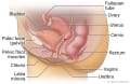
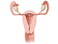
A pelvic ultrasound is a test that uses sound waves to make a picture of the organs and structures in the lower belly (pelvis).
This test looks at the bladder and:
Organs and structures that are solid and uniform (such as the uterus, ovaries, or prostate gland) or that are fluid-filled (such as the bladder) show up clearly on a pelvic ultrasound. Bones may block other organs from being seen. Air-filled organs, such as the intestines, can make the image less clear.
Pelvic ultrasound can be done in several ways.
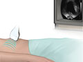
- Transabdominal ultrasound.
-
A small handheld device called a transducer is passed back and forth over the lower belly.
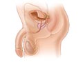
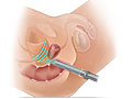
- Transrectal ultrasound.
-
The transducer is shaped to fit into the rectum. A transrectal ultrasound is the most common test to look at the male pelvic organs, such as the prostate and seminal vesicles. The test may also be done to look for rectal problems in men or women.
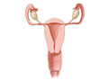
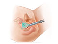
- Transvaginal ultrasound.
-
The transducer is shaped to fit into a woman's vagina. A woman may have both transabdominal and transvaginal ultrasounds to look at the whole pelvic area.
- Transperineal and translabial ultrasound.
- Both of these types of ultrasound are used on the outside of the genital area to look for urinary and pelvic problems.
In all of these ultrasounds, the transducer sends the reflected sound waves to a computer, which makes them into a picture that is shown on a video screen. Ultrasound pictures or videos may be saved as a permanent record.