Test Overview
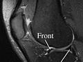
Magnetic resonance imaging (MRI) is a test done with a large machine that uses a magnetic field and pulses of radio wave energy to make pictures of the knee. Muscles, ligaments, cartilage, and other joint structures are often best seen with an MRI. In many cases an MRI gives information about structures in the body that cannot be seen as well with an X-ray, ultrasound, or CT scan.
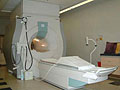
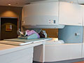
For an MRI test, you are placed inside the magnet so that your knee is inside the strong magnetic field. An MRI can find changes in the structure of organs or other tissues. It also can find tissue damage or disease, such as infection or a tumor. Pictures from an MRI scan are digital images that can be saved and stored on a computer for further study. The images also can be reviewed remotely, such as in a clinic or an operating room. Photographs or films of selected pictures can also be made.
In some cases, a contrast material may be used during the MRI scan to show certain structures more clearly in the pictures. The contrast material may be used to check blood flow, find some types of tumors, and show areas of inflammation or infection. The contrast material may be put in a vein (I.V.) in your arm or directly into your knee.
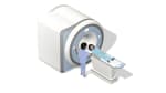
There are two main types of MRI—the closed MRI machine and the open MRI machine.