Test Overview
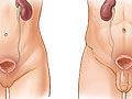
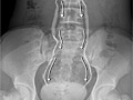
An intravenous pyelogram (IVP) is an X-ray test that provides pictures of the kidneys, the bladder, the ureters, and the urethra (urinary tract). An IVP can show the size, shape, and position of the urinary tract, and it can evaluate the collecting system inside the kidneys.
During IVP, a dye called contrast material is injected into a vein in your arm. A series of X-ray pictures is then taken at timed intervals.
IVP is commonly done to identify diseases of the urinary tract, such as kidney stones, tumors, or infection. It is also used to look for problems with the structure of the urinary tract that were present from birth (congenital).
An ultrasound or a computed tomography (CT) scan may be combined with an IVP if more details about the urinary tract are needed. A computed tomography intravenous pyelogram (CT/IVP) is usually done to look for the cause of blood in the urine.