Test Overview
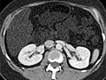
A computed tomography (CT) scan uses X-rays to make detailed pictures of structures inside of the body.
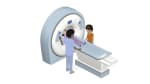
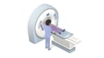
During the test, you will lie on a table that is attached to the CT scanner, which is a large doughnut-shaped machine. The CT scanner sends X-rays through the body area being studied. Each rotation of the scanner provides a picture of a thin slice of the organ or area. All of the pictures are saved as a group on a computer. They also can be printed.
In some cases, a dye called contrast material may be used. It may be put in a vein (I.V.) in your arm. Or it may be placed into other parts of your body (such as the rectum or a joint) to see those areas better. For some types of CT scans, you drink the dye. The dye makes structures and organs easier to see on the CT pictures.
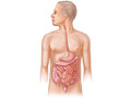
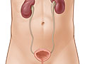
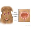
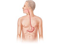
A CT scan can be used to study all parts of your body, such as the chest, belly, pelvis, or an arm or leg. It can take pictures of body organs, such as the liver, pancreas, intestines, kidneys, bladder, adrenal glands, lungs, and heart. It also can study blood vessels, bones, and the spinal cord.