Test Overview
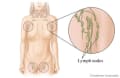
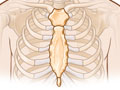
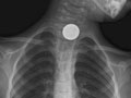
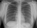
A chest X-ray is a picture of the chest that shows your heart, lungs, airway, blood vessels, and lymph nodes. A chest X-ray also shows the bones of your spine and chest, including your breastbone, your ribs, your collarbone, and the upper part of your spine. A chest X-ray is the most common imaging test or X-ray used to find problems inside the chest.
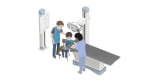
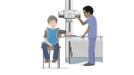
A chest X-ray can help find some problems with the organs and structures inside the chest. Usually two pictures are taken, one from the back of the chest and another from the side. In an emergency when only one X-ray picture is taken, a front view is usually done. Doctors may not always get the information they need from a chest X-ray to find the cause of a problem. If the results from a chest X-ray are not normal or do not give enough information about the chest problem, more specific X-rays or other tests may be done, such as a computed tomography (CT) scan, an ultrasound, an echocardiogram, or an MRI scan.