Test Overview
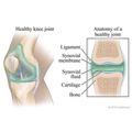
An arthrogram is a test using X-rays to obtain a series of pictures of a joint after a contrast material (such as a dye, water, air, or a combination of these) has been injected into the joint. This allows your doctor to see the soft tissue structures of your joint, such as tendons, ligaments, muscles, cartilage, and your joint capsule. These structures are not seen on a plain X-ray without contrast material. A special type of X-ray, called fluoroscopy, is used to take pictures of the joint.
An arthrogram is used to check a joint to find out what is causing your symptoms or problem with your joint. An arthrogram may be more useful than a regular X-ray because it shows the surface of soft tissues lining the joint as well as the joint bones. A regular X-ray only shows the bones of the joint. This test can be done on your hip, knee, ankle, shoulder, elbow, wrist, or jaw (temporomandibular joint).
Other tests, such as magnetic resonance imaging (MRI) and computed tomography (CT), give different information about a joint. They may be used with an arthrogram or when an arthrogram does not give a clear picture of the joint.