Test Overview
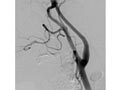
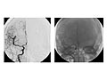
An angiogram of the head and neck is an X-ray test that uses a special dye and camera (fluoroscopy) to take pictures of the blood flow in the blood vessels of the head and neck. An angiogram of the neck (carotid angiogram) can be used to look at the large arteries in the neck that lead to the brain. An angiogram of the head (cerebral angiogram) can be used to look at the veins or the four arteries (four-vessel study) carrying blood to the brain.
During an angiogram, a thin, soft tube called a catheter is placed into a blood vessel in the groin or just above the elbow. The catheter is guided to the head and neck area. Then an iodine dye (contrast material) is injected into the vessel to make the area show clearly on the X-ray pictures. The angiogram pictures can be made into regular X-ray films or stored as digital pictures in a computer.
An angiogram can find a bulge in a blood vessel (aneurysm). It can also show narrowing or a blockage in a blood vessel that slows or stops blood flow. An abnormal pattern of blood vessels (arteriovenous [AV] malformation) or abnormal vessels near a tumor can be seen.
A magnetic resonance angiogram (MRA) or computed tomography angiogram (CTA) may be an option instead of a standard angiogram. Each of these tests is less invasive than an angiogram. Some MRA tests and all CTA tests require an injection of dye. A CTA also involves radiation exposure.
Current as of: July 31, 2024
© 2017-2025 Healthwise, Incorporated. This information does not replace the advice of a doctor.