


General Information About Thymoma and Thymic Carcinoma
Thymoma and thymic carcinoma are diseases in which malignant (cancer) cells form in the thymus.
Thymoma and thymic carcinoma are two types of cancer that can form in the cells that cover the outside surface of the thymus. The thymus is a small organ in the upper chest under the breastbone. It is part of the lymph system and makes white blood cells, called lymphocytes, that help fight infection. These cancers usually form between the lungs in the front part of the chest and are often found during a chest x-ray that is done for another reason.
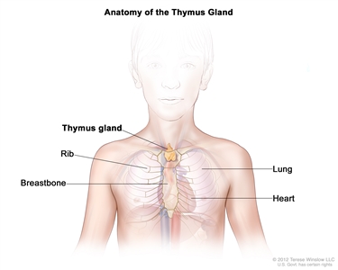
Anatomy of the thymus gland. The thymus gland is a small organ that lies in the upper chest under the breastbone. It makes white blood cells, called lymphocytes, which protect the body against infections.
Even though thymoma and thymic carcinoma form in the same type of cell, they act differently:
- Thymoma. The cancer cells look a lot like the normal cells of the thymus, grow slowly, and rarely spread beyond the thymus. A thymoma may become a thymic carcinoma over time.
- Thymic carcinoma. The cancer cells do not look like the normal cells of the thymus, grow more quickly, and are more likely to spread to other parts of the body.
Other types of tumors, such as lymphoma or germ cell tumors, may form in the thymus, but they are not considered to be thymoma or thymic carcinoma.
Signs and symptoms of thymoma and thymic carcinoma include coughing and trouble breathing.
These and other signs and symptoms may be caused by thymoma and thymic carcinoma or by other conditions.
Check with your child's doctor if your child has any of the following:
- Coughing.
- Trouble breathing.
- Pain or a tight feeling in the chest.
- Trouble swallowing.
- Hoarseness.
- Fever.
- Weight loss.
- Superior vena cava syndrome.
Children with thymoma or thymic carcinoma may also have other health problems.
Children who have thymoma or thymic carcinoma may also have one of the following immune system diseases or hormone disorders:
- Myasthenia gravis.
- Pure red cell aplasia.
- Hypogammaglobulinemia.
- Nephrotic syndrome.
- Scleroderma.
- Dermatomyositis.
- Lupus.
- Rheumatoid arthritis.
- Thyroiditis.
- Hyperthyroidism.
- Addison disease.
- Panhypopituitarism.
Tests that examine the thymus and chest are used to help diagnose thymoma and thymic carcinoma.
The following tests and procedures may be used:
- Physical exam and health history: An exam of the body to check general signs of health, including checking for signs of disease, such as lumps or anything else that seems unusual. A history of the patient's health habits and past illnesses and treatments will also be taken.
- Chest x-ray: An x-ray of the organs and bones inside the chest. An x-ray is a type of energy beam that can go through the body and onto film, making a picture of areas inside the body.
- CT scan (CAT scan): A procedure that makes a series of detailed pictures of areas inside the body, taken from different angles. The pictures are made by a computer linked to an x-ray machine. A dye may be injected into a vein or swallowed to help the organs or tissues show up more clearly. This procedure is also called computed tomography, computerized tomography, or computerized axial tomography.
- MRI (magnetic resonance imaging): A procedure that uses a magnet, radio waves, and a computer to make a series of detailed pictures of areas inside the body. This procedure is also called nuclear magnetic resonance imaging (NMRI).
- PET scan (positron emission tomography scan): A procedure to find malignant tumor cells in the body. A small amount of radioactive glucose (sugar) is injected into a vein. The PET scanner rotates around the body and makes a picture of where glucose is being used in the body. Malignant tumor cells show up brighter in the picture because they are more active and take up more glucose than normal cells do.
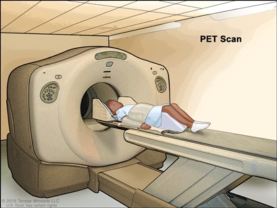
Positron emission tomography (PET) scan. The child lies on a table that slides through the PET scanner. The head rest and white strap help the child lie still. A small amount of radioactive glucose (sugar) is injected into the child's vein, and a scanner makes a picture of where the glucose is being used in the body. Cancer cells show up brighter in the picture because they take up more glucose than normal cells do. - Biopsy: The removal of cells or tissues so they can be viewed under a microscope by a pathologist to check for signs of cancer.
Certain factors affect prognosis (chance of recovery).
The prognosis depends on the following:
- Whether the cancer is thymoma or thymic carcinoma.
- Whether the cancer has spread to nearby areas or other parts of the body.
- Whether the cancer was completely removed by surgery.
- Whether the cancer is newly diagnosed or has recurred (come back) after treatment.
The prognosis is better when the cancer has not spread to other parts of the body. Childhood thymoma is usually diagnosed before the tumor has spread.