


General Information About Childhood Pheochromocytoma and Paraganglioma
Pheochromocytoma forms in the adrenal gland.
Pheochromocytoma forms in the adrenal gland. There are two adrenal glands, one on top of each kidney in the back of the upper abdomen. Each adrenal gland has two parts. The outer layer of the adrenal gland is the adrenal cortex. The center of the adrenal gland is the adrenal medulla. Pheochromocytoma is a tumor of the adrenal medulla.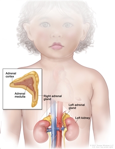
Anatomy of the adrenal gland. There are two adrenal glands, one on top of each kidney. The outer part of each gland is the adrenal cortex and the inner part is the adrenal medulla.
The adrenal glands make important hormones called catecholamines. Adrenaline (epinephrine) and noradrenaline (norepinephrine) are two types of catecholamines that help control heart rate, blood pressure, blood sugar, and the way the body reacts to stress. Some pheochromocytomas release extra adrenaline and noradrenaline into the blood and cause symptoms.
Paraganglioma forms in nerve tissue near the carotid artery, along nerve pathways in the head and neck, and in other parts of the body.
Some paragangliomas make extra catecholamines called adrenaline and noradrenaline. The release of extra adrenaline and noradrenaline into the blood may cause symptoms.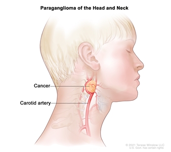
A paraganglioma is a rare tumor that often forms in nerve tissue near the carotid artery. It may also form along nerve pathways in the head and neck and in other parts of the body.
Pheochromocytoma and paraganglioma may be benign (not cancer) or malignant (cancer).
About half of all children with pheochromocytoma or paraganglioma have malignant pheochromocytoma or paraganglioma. This means that the tumor cells have spread to other parts of the body. Benign and malignant pheochromocytoma and paraganglioma require treatment because they can cause severe or life-threatening heart problems and affect many body functions.
Inheriting certain gene mutations (changes) increases the risk of pheochromocytoma and paraganglioma.
Anything that increases your chance of getting a disease is called a risk factor. Having a risk factor does not mean that you will get cancer; not having risk factors doesn't mean that you will not get cancer. Talk with your child's doctor if you think your child may be at risk.
Having any of the following inherited syndromes or gene changes increases risk of pheochromocytoma and paraganglioma:
- Multiple endocrine neoplasia type 1 (MEN1) syndrome (tumors in the parathyroid gland, pituitary gland, or islet cells in the pancreas, and rarely, pheochromocytoma).
- Multiple endocrine neoplasia type 2A syndrome (pheochromocytoma, medullary thyroid cancer, and parathyroid gland disease).
- Multiple endocrine neoplasia type 2B syndrome (pheochromocytoma, medullary thyroid cancer, parathyroid hyperplasia, and other conditions).
- von Hippel-Lindau disease (VHL) (pheochromocytoma, paraganglioma, hemangioblastoma, clear cell renal carcinoma, pancreatic neuroendocrine tumors, and other conditions).
- Neurofibromatosis type 1 (NF1) (neurofibromas, brain tumors, pheochromocytoma, and other conditions).
- Carney-Stratakis dyad (paraganglioma and gastrointestinal stromal tumor [GIST]).
- Carney triad (paraganglioma, GIST, and pulmonary chondroma).
- Familial pheochromocytoma or paraganglioma.
More than half of the children and adolescents diagnosed with pheochromocytoma or paraganglioma have an inherited syndrome or gene change that increases the risk of cancer. Genetic counseling (a discussion with a trained professional about inherited diseases) and testing is an important part of the treatment plan.
Signs and symptoms of pheochromocytoma and paraganglioma occur if too much adrenaline or noradrenaline is released into the blood.
Some tumors do not make extra adrenaline or noradrenaline and do not cause symptoms. These tumors may be found when a lump forms in the neck or when a test is done for another reason. Signs and symptoms of pheochromocytoma and paraganglioma occur when too much adrenaline or noradrenaline is released into the blood. These and other signs and symptoms may be caused by pheochromocytoma, paraganglioma, or other conditions.
Check with your child's doctor if your child has any of the following:
- High blood pressure.
- Headache.
- Heavy sweating for no known reason.
- A strong, fast, or irregular heartbeat.
- Feeling shaky.
- Being extremely pale.
- Dizziness.
- Being irritable or nervous.
These signs and symptoms may come and go, but high blood pressure is more likely to occur for long periods of time in young patients. These signs and symptoms may also occur with physical activity, injury, anesthesia, surgery to remove the tumor, eating foods such as chocolate and cheese, or while passing urine (if the tumor is in the bladder).
Tests and procedures used to diagnose and stage pheochromocytoma and paraganglioma depend on the patient's signs and symptoms and family history.
Tests are done to diagnose and stage cancer. After cancer is diagnosed, more tests are done to find out if cancer cells have spread to nearby areas or to other parts of the body. This process is called staging.
The following tests and procedures may be used:
- Physical exam and health history: An exam of the body to check general signs of health, including checking for signs of disease, such as lumps or anything else that seems unusual. A history of the patient's health habits and past illnesses and treatments will also be taken.
- Plasma -free metanephrines test: A blood test that measures the amount of metanephrines in the blood. Metanephrines are substances that are made when the body breaks down adrenaline or noradrenaline. Pheochromocytomas and paragangliomas can make large amounts of adrenaline and noradrenaline and cause high levels of metanephrines in both the blood and urine.
- Blood catecholamine studies: A procedure in which a blood sample is checked to measure the amount of certain catecholamines (adrenaline or noradrenaline) released into the blood. Substances caused by the breakdown of these catecholamines are also measured. An unusual (unusual higher or lower than normal) amount of a substance can be a sign of disease in the organ or tissue that makes it. Higher than normal amounts may be a sign of pheochromocytoma or paraganglioma.
- Twenty-four-hour urine test: A test in which urine is collected for 24 hours to measure the amounts of catecholamines (adrenaline or noradrenaline) or metanephrines in the urine. Substances caused by the breakdown of these catecholamines are also measured. An unusual (higher than normal) amount of a substance can be a sign of disease in the organ or tissue that makes it. Higher than normal amounts may be a sign of pheochromocytoma or paraganglioma.
- PET scan: A procedure to find malignant tumor cells in the body. A small amount of radioactive glucose (sugar) is injected into a vein. The PET scanner rotates around the body and makes a picture of where glucose is being used in the body. Malignant tumor cells show up brighter in the picture because they are more active and take up more glucose than normal cells do.
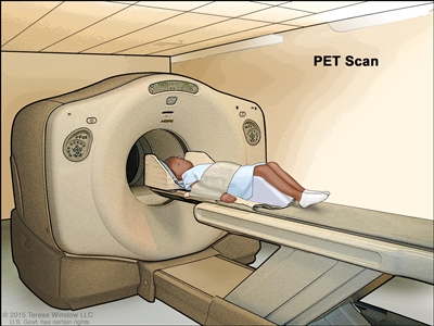
Positron emission tomography (PET) scan. The child lies on a table that slides through the PET scanner. The head rest and white strap help the child lie still. A small amount of radioactive glucose (sugar) is injected into the child's vein, and a scanner makes a picture of where the glucose is being used in the body. Cancer cells show up brighter in the picture because they take up more glucose than normal cells do. - CT scan (CAT scan): A procedure that makes a series of detailed pictures of areas inside the body, taken from different angles. The pictures are made by a computer linked to an x-ray machine. A dye may be injected into a vein or swallowed to help the organs or tissues show up more clearly. This procedure is also called computed tomography, computerized tomography, or computerized axial tomography.
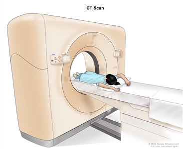
Computed tomography (CT) scan. The child lies on a table that slides through the CT scanner, which takes a series of detailed x-ray pictures of areas inside the body. - MRI (magnetic resonance imaging): A procedure that uses a magnet, radio waves, and a computer to make a series of detailed pictures of areas inside the body. This procedure is also called nuclear magnetic resonance imaging (NMRI).
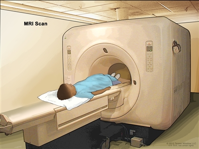
Magnetic resonance imaging (MRI) scan. The child lies on a table that slides into the MRI machine, which takes a series of detailed pictures of areas inside the body. The positioning of the child on the table depends on the part of the body being imaged. - MIBG scan: A procedure used to find neuroendocrine tumors, such as pheochromocytoma and paraganglioma. A very small amount of a substance called radioactive MIBG is injected into a vein and travels through the bloodstream. Neuroendocrine tumor cells take up the radioactive MIBG and are detected by a scanner. Scans may be taken over 1-3 days. An iodine solution may be given before or during the test to keep the thyroid gland from absorbing too much of the MIBG.
- Somatostatin receptor scintigraphy: A type of radionuclide scan that may be used to find tumors. A very small amount of radioactive octreotide (a hormone that attaches to tumors) is injected into a vein and travels through the blood. The radioactive octreotide attaches to the tumor and a special camera that detects radioactivity is used to show where the tumors are in the body. This procedure is also called octreotide scan and SRS.
- Genetic testing: A laboratory test in which cells or tissue are analyzed to look for changes in genes or chromosomes. These changes may be a sign that a person has or is at risk of having a specific disease or condition. The following are genes that might be tested for in children with pheochromocytoma or paraganglioma: VHL, NF1, RET, SDHD, SDHB, SDHA, MAX, and TMEM127 genes.
Certain factors affect prognosis (chance of recovery) and treatment options.
Prognosis and treatment options depend on the following:
- Whether the cancer has just been diagnosed or has recurred (come back).