


General Information About Childhood Non-Hodgkin Lymphoma
Childhood non-Hodgkin lymphoma is a disease in which malignant (cancer) cells form in the lymph system, which is a part of the body's immune system.
The immune system helps protect the body from infection and disease.
The lymph system is made up of the following:
- Lymph: Colorless, watery fluid that travels through the lymph vessels and carries lymphocytes (white blood cells).
- B lymphocytes, also called B cells, make antibodies to help fight infection. Most types of non-Hodgkin lymphoma begin in B lymphocytes.
- T lymphocytes, also called T cells, help B lymphocytes make the antibodies that help fight infection.
- Natural killer cells, also called NK cells, attack cancer cells and viruses.
- Lymph vessels: A network of thin tubes that collect lymph from different parts of the body and return it to the bloodstream.
- Lymph nodes: Small, bean-shaped structures that filter lymph and store white blood cells that help fight infection and disease. Lymph nodes are found along a network of lymph vessels throughout the body. Groups of lymph nodes are found in the neck, underarm, mediastinum (the area between the lungs), abdomen, pelvis, and groin.
- Spleen: An organ that makes lymphocytes, stores red blood cells and lymphocytes, filters the blood, and destroys old blood cells. The spleen is on the left side of the abdomen near the stomach.
- Thymus: An organ in which T lymphocytes mature and multiply. The thymus is in the chest behind the breastbone.
- Tonsils: Two small masses of lymph tissue at the back of the throat. There is one tonsil on each side of the throat.
- Bone marrow: The soft, spongy tissue in the center of certain bones, such as the hip bone and breastbone. White blood cells, red blood cells, and platelets are made in the bone marrow.
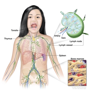
The lymph system is part of the body's immune system and is made up of tissues and organs that help protect the body from infection and disease. These include the tonsils, thymus, spleen, bone marrow, lymph vessels, and lymph nodes. Lymph (clear, watery fluid) and lymphocytes (white blood cells) travel through the lymph vessels and into the lymph nodes where the lymphocytes destroy harmful substances. The lymph enters the bloodstream through a large vein near the heart.
Lymph tissue is also found in other parts of the body such as the stomach, thyroid gland, brain, and skin.
Non-Hodgkin lymphoma can begin in B lymphocytes, T lymphocytes, or natural killer cells.
There are two general types of lymphomas: Hodgkin lymphoma and non-Hodgkin lymphoma. This summary is about the treatment of childhood non-Hodgkin lymphoma. For information about the treatment of childhood Hodgkin lymphoma, see Childhood Hodgkin Lymphoma Treatment.
Treatment of non-Hodgkin lymphoma is different for children and adults. For information about treatment of adults, see the following:
- Adult Non-Hodgkin Lymphoma
- Primary CNS Lymphoma Treatment
- Mycosis Fungoides (Including Sezary Syndrome) Treatment
There are three major types of childhood non-Hodgkin lymphoma.
The type of lymphoma is determined by how the cells look under a microscope. The three major types of childhood non-Hodgkin lymphoma are:
Aggressive mature B-cell non-Hodgkin lymphoma
Aggressive mature B-cell non-Hodgkin lymphomas include:
- Burkitt lymphoma/leukemia: Burkitt lymphoma and Burkitt leukemia are different forms of the same disease. Burkitt lymphoma/leukemia is an aggressive (fast-growing) disorder of B lymphocytes that is most common in children and young adults. It may form in the abdomen, Waldeyer's ring, testicles, bone, bone marrow, skin, central nervous system (CNS), or head and neck. Burkitt leukemia may start in the lymph nodes as Burkitt lymphoma and then spread to the blood and bone marrow, or it may start in the blood and bone marrow without forming in the lymph nodes first.
Both Burkitt leukemia and Burkitt lymphoma have been linked to infection with the Epstein-Barr virus (EBV), although EBV infection is more likely to occur in patients in Africa than in the United States. Burkitt lymphoma/leukemia is more common in White people. Burkitt lymphoma/leukemia is diagnosed when a sample of tissue is checked and a certain change to the MYCgene is found.
- Diffuse large B-cell lymphoma: Diffuse large B-cell lymphoma is the most common type of non-Hodgkin lymphoma. It is a type of B-cell non-Hodgkin lymphoma that grows quickly in the lymph nodes. The spleen, liver, bone marrow, or other organs are also often affected. Diffuse large B-cell lymphoma occurs more often in adolescents than in children.
- Primary mediastinal B-cell lymphoma: A type of lymphoma that develops from B cells in the mediastinum (the area between the lungs). It may spread to nearby organs including the lungs and the sac around the heart. It may also spread to lymph nodes and distant organs including the kidneys. Primary mediastinal B-cell lymphoma occurs more often in older adolescents than in children.
Lymphoblastic lymphoma
Lymphoblastic lymphoma is a type of lymphoma that mainly affects T-cell lymphocytes. It usually forms in the mediastinum (the area between the lungs). This causes trouble breathing, wheezing, trouble swallowing, or swelling of the head and neck. It may spread to lymph nodes, bone, bone marrow, skin, the CNS, abdominal organs, and other areas. Lymphoblastic lymphoma is a lot like acute lymphoblastic leukemia (ALL).
Anaplastic large cell lymphoma
Anaplastic large cell lymphoma is a type of lymphoma that mainly affects T-cell lymphocytes. It usually forms in the lymph nodes, skin, or bone, and sometimes forms in the gastrointestinal tract, lung, tissue that covers the lungs, and muscle. Patients with anaplastic large cell lymphoma have a receptor, called CD30, on the surface of their T cells. In many children, anaplastic large cell lymphoma is marked by changes in the ALK gene that makes a protein called anaplastic lymphoma kinase. A pathologist checks for these cell and gene changes to help diagnose anaplastic large cell lymphoma.
Some types of non-Hodgkin lymphoma are rare in children.
Some types of childhood non-Hodgkin lymphoma are less common. These include:
- Pediatric-type follicular lymphoma: In children, follicular lymphoma occurs mainly in males. It is more likely to be found in one area and does not spread to other places in the body. It usually forms in the tonsils and lymph nodes in the neck, but may also form in the testicles, kidney, gastrointestinal tract, and salivary gland.
- Marginal zone lymphoma: Marginal zone lymphoma is a type of lymphoma that tends to grow and spread slowly and is usually found at an early stage. It may be found in the lymph nodes or in areas outside the lymph nodes. Marginal zone lymphoma found outside the lymph nodes in children is called mucosa-associated lymphoid tissue (MALT) lymphoma. MALT may be linked to Helicobacter pylori infection of the gastrointestinal tract and Chlamydophila psittaci infection of the conjunctival membrane which lines the eye. Marginal zone lymphoma is rare in children but common in adults.
- Primary central nervous system (CNS) lymphoma: Primary CNS lymphoma is extremely rare in children.
- Peripheral T-cell lymphoma: Peripheral T-cell lymphoma is an aggressive (fast-growing) non-Hodgkin lymphoma that begins in mature T lymphocytes. Other types of peripheral T-cell lymphoma include mature T-cell/natural killer-cell lymphoma, extranodal NK/T-cell lymphoma, and gamma-delta hepatosplenic T-cell lymphoma. Peripheral T-cell lymphoma is rare in children.
- Cutaneous T-cell lymphoma: Cutaneous T-cell lymphoma begins in the skin and can cause the skin to thicken or form a tumor. It is very rare in children but is more common in adolescents and young adults. There are different types of cutaneous T-cell lymphoma, such as cutaneous anaplastic large cell lymphoma, subcutaneous panniculitis-like T-cell lymphoma, gamma-delta T-cell lymphoma, and mycosis fungoides. Mycosis fungoides rarely occurs in children and adolescents.
Having a weakened immune system increases the risk of NHL in children.
Anything that increases a person's risk of getting a disease is called a risk factor. Not every child with one or more of these risk factors will develop NHL, and it will develop in some children who don't have any known risk factors. Talk with your child's doctor if you think your child may be at risk.
Some of the types of immune system problems that have been linked with a higher risk of childhood NHL include the following:
- Being infected with the Epstein-Barr virus or human immunodeficiency virus (HIV).
- Having a weakened immune system after a transplant or from medicines given after a transplant.
- Having certain inherited diseases (such as DNA repair defect syndromes which include ataxia-telangiectasia, Nijmegen breakage syndrome, and constitutional mismatch repair deficiency).
- Past treatment for cancer.
If lymphoma or lymphoproliferative disease is linked to a weakened immune system from certain inherited diseases, HIV infection, a transplant or medicines given after a transplant, the condition is called lymphoproliferative disease associated with immunodeficiency. The different types of lymphoproliferative disease associated with immunodeficiency include:
- Lymphoproliferative disease associated with primary immunodeficiency.
- HIV-associated non-Hodgkin lymphoma.
- Post-transplant lymphoproliferative disease.
- Lymphoproliferative disease from chemotherapy.
Signs of childhood non-Hodgkin lymphoma include breathing problems and swollen lymph nodes.
These and other signs may be caused by childhood non-Hodgkin lymphoma or by other conditions. Check with a doctor if your child has any of the following:
- Trouble breathing.
- Wheezing.
- Coughing.
- High-pitched breathing sounds.
- Swelling of the head, neck, upper body, or arms.
- Trouble swallowing.
- Painless swelling of the lymph nodes in the neck, underarm, stomach, or groin.
- Painless lump or swelling in a testicle.
- Fever for no known reason.
- Weight loss for no known reason.
- Drenching night sweats.
- Pain or swelling in the abdomen.
Tests that examine the body and lymph system are used to diagnose childhood non-Hodgkin lymphoma.
In addition to asking about your child's personal and family health history and doing a physical exam, your child's doctor may perform the following tests and procedures:
- Blood chemistry studies: A procedure in which a blood sample is checked to measure the amounts of certain substances released into the blood by organs and tissues in the body, including electrolytes, lactate dehydrogenase (LDH), uric acid, blood urea nitrogen (BUN), creatinine, and liver function values. An unusual (higher or lower than normal) amount of a substance can be a sign of disease.
- Liver function tests: A procedure in which a blood sample is checked to measure the amounts of certain substances released into the blood by the liver. A higher-than-normal amount of a substance can be a sign of cancer.
- CT scan (CAT scan): A procedure that makes a series of detailed pictures of areas inside the body, such as the neck, chest, abdomen, and pelvis, taken from different angles. The pictures are made by a computer linked to an x-ray machine. A dye may be injected into a vein or swallowed to help the organs or tissues show up more clearly. This procedure is also called computed tomography, computerized tomography, or computerized axial tomography.
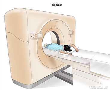
Computed tomography (CT) scan. The child lies on a table that slides through the CT scanner, which takes a series of detailed x-ray pictures of areas inside the body. - PET scan (positron emission tomography scan): A procedure to find malignant tumor cells in the body. A small amount of radioactive glucose (sugar) is injected into a vein. The PET scanner rotates around the body and makes a picture of where glucose is being used in the body. Malignant tumor cells show up brighter in the picture because they are more active and take up more glucose than normal cells do. Sometimes a PET scan and a CT scan are done at the same time. If there is any cancer, this increases the chance that it will be found.
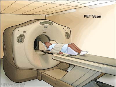
Positron emission tomography (PET) scan. The child lies on a table that slides through the PET scanner. The head rest and white strap help the child lie still. A small amount of radioactive glucose (sugar) is injected into the child's vein, and a scanner makes a picture of where the glucose is being used in the body. Cancer cells show up brighter in the picture because they take up more glucose than normal cells do. - MRI (magnetic resonance imaging): A procedure that uses a magnet, radio waves, and a computer to make a series of detailed pictures of areas inside the body. This procedure is also called nuclear magnetic resonance imaging (NMRI).
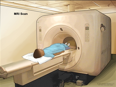
Magnetic resonance imaging (MRI) scan. The child lies on a table that slides into the MRI machine, which takes a series of detailed pictures of areas inside the body. The positioning of the child on the table depends on the part of the body being imaged. - Lumbar puncture: A procedure used to collect cerebrospinal fluid (CSF) from the spinal column. This is done by placing a needle between two bones in the spine and into the CSF around the spinal cord and removing a sample of the fluid. The sample of CSF is checked under a microscope for signs that the cancer has spread to the brain and spinal cord. This procedure is also called an LP or spinal tap.
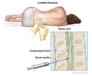
Lumbar puncture. A patient lies in a curled position on a table. After a small area on the lower back is numbed, a spinal needle (a long, thin needle) is inserted into the lower part of the spinal column to remove cerebrospinal fluid (CSF, shown in blue). The fluid may be sent to a laboratory for testing. - Chest x-ray: An x-ray of the organs and bones inside the chest. An x-ray is a type of energy beam that can go through the body and onto film, making a picture of areas inside the body.
- Ultrasound exam: A procedure in which high-energy sound waves (ultrasound) are bounced off internal tissues or organs and make echoes. The echoes form a picture of body tissues called a sonogram.
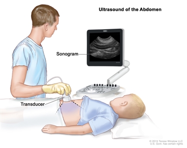
Abdominal ultrasound. An ultrasound transducer connected to a computer is pressed against the skin of the abdomen. The transducer bounces sound waves off internal organs and tissues to make echoes that form a sonogram (computer picture).
A biopsy is done to diagnose childhood non-Hodgkin lymphoma.
Cells and tissues are removed during a biopsy so they can be viewed under a microscope by a pathologist to check for cancer cells. Because treatment depends on the type of non-Hodgkin lymphoma, biopsy samples should be checked by a pathologist who has experience in diagnosing childhood non-Hodgkin lymphoma.
One of the following types of biopsies may be done:
- Excisional biopsy: The removal of an entire lymph node or lump of tissue.
- Incisional biopsy: The removal of part of a lump, lymph node, or sample of tissue.
- Core needle biopsy: The removal of tissue or part of a lymph node using a wide needle.
- Fine-needle aspiration (FNA) biopsy: The removal of tissue or part of a lymph node using a thin needle.
The procedure used to remove the sample of tissue depends on where the tumor is in the body:
- Bone marrow aspiration and biopsy: The removal of bone marrow and a small piece of bone by inserting a hollow needle into the hipbone or breastbone.
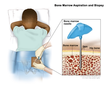
Bone marrow aspiration and biopsy. After a small area of skin is numbed, a bone marrow needle is inserted into the child's hip bone. Samples of blood, bone, and bone marrow are removed for examination under a microscope. - Mediastinoscopy: A surgical procedure to look at the organs, tissues, and lymph nodes between the lungs for abnormal areas. An incision (cut) is made at the top of the breastbone and a mediastinoscope is inserted into the chest. A mediastinoscope is a thin, tube-like instrument with a light and a lens for viewing. It also has a tool to remove tissue or lymph node samples, which are checked under a microscope for signs of cancer.
- Anterior mediastinotomy: A surgical procedure to look at the organs and tissues between the lungs and between the breastbone and heart for abnormal areas. An incision (cut) is made next to the breastbone and a mediastinoscope is inserted into the chest. A mediastinoscope is a thin, tube-like instrument with a light and a lens for viewing. It also has a tool to remove tissue or lymph node samples, which are checked under a microscope for signs of cancer. This is also called the Chamberlain procedure.
- Thoracentesis: The removal of fluid from the space between the lining of the chest and the lung, using a needle. A pathologist views the fluid under a microscope to look for cancer cells.
If cancer is found, the following tests may be done to study the cancer cells:
- Immunohistochemistry: A laboratory test that uses antibodies to check for certain antigens (markers) in a sample of a patient's tissue. The antibodies are usually linked to an enzyme or a fluorescent dye. After the antibodies bind to a specific antigen in the tissue sample, the enzyme or dye is activated, and the antigen can then be seen under a microscope. This type of test is used to help diagnose cancer and to help tell one type of cancer from another type of cancer.
- Flow cytometry: A laboratory test that measures the number of cells in a sample, the percentage of live cells in a sample, and certain characteristics of the cells, such as size, shape, and the presence of tumor (or other) markers on the cell surface. The cells from a sample of a patient's blood, bone marrow, or other tissue are stained with a fluorescent dye, placed in a fluid, and then passed one at a time through a beam of light. The test results are based on how the cells that were stained with the fluorescent dye react to the beam of light. This test is used to help diagnose and manage certain types of cancers, such as leukemia and lymphoma.
- Cytogenetic analysis: A laboratory test in which the chromosomes of cells in a sample of blood or bone marrow are counted and checked for any changes, such as broken, missing, rearranged, or extra chromosomes. Changes in certain chromosomes may be a sign of cancer. Cytogenetic analysis is used to help diagnose cancer, plan treatment, or find out how well treatment is working.
- FISH (fluorescence in situ hybridization): A laboratory test used to look at and count genes or chromosomes in cells and tissues. Pieces of DNA that contain fluorescent dyes are made in the laboratory and added to a sample of a patient's cells or tissues. When these dyed pieces of DNA attach to certain genes or areas of chromosomes in the sample, they light up when viewed under a fluorescent microscope. The FISH test is used to help diagnose cancer and help plan treatment.
- Immunophenotyping: A laboratory test that uses antibodies to identify cancer cells based on the types of antigens or markers on the surface of the cells. This test is used to help diagnose specific types of lymphoma.
Certain factors affect prognosis (chance of recovery) and treatment options.
The prognosis and treatment options depend on:
- The type of lymphoma.
- Where the tumor is in the body when the tumor is diagnosed.
- The stage of the cancer.
- Whether there are certain changes in the chromosomes.
- The type of initial treatment.
- Whether the lymphoma responded to initial treatment.
- The patient's age and general health.