


General Information About Childhood Mesothelioma
Mesothelioma is a disease in which malignant (cancer) cells form in the thin layer of tissue that covers organs in the chest or abdomen.
Malignant mesothelioma is a disease in which malignant (cancer) cells are found in one or more of the following:
- Pleura: A thin layer of tissue that lines the chest cavity and covers the lungs.
- Peritoneum: A thin layer of tissue that lines the abdomen and covers most of the organs in the abdomen.
- Pericardium: A thin layer of tissue that surrounds the heart.
The tumors often spread over the surface of organs without spreading into the organ. They may spread to nearby lymph nodes or in other parts of the body. Malignant mesothelioma may also form in the testicles, but this is rare.

Malignant mesothelioma forms in the tissue that lines the chest or abdomen, including the pleura (the tissue that lines the chest cavity and covers the lungs), the pericardium (the tissue that surrounds the heart), and the peritoneum (the tissue that lines the abdomen and covers most of the organs in the abdomen). Malignant mesothelioma may also form in the testicles, but this is rare.
Treatment with radiation therapy increases the risk of childhood mesothelioma.
Anything that increases your chance of getting a disease is called a risk factor. Having a risk factor does not mean that you will get cancer; not having risk factors doesn't mean that you will not get cancer. Talk with your child's doctor if you think your child may be at risk.
Treatment for an earlier cancer, especially radiation therapy, increases the risk of mesothelioma in children.
In adults, mesothelioma is strongly linked to being exposed to asbestos, which has been used in the building and textile industries. In children, there is little information about the risk of developing mesothelioma after being exposed to asbestos.
Signs and symptoms of mesothelioma include trouble breathing and pain in the chest or abdomen.
In children, these and other signs and symptoms may be caused by mesothelioma or by other conditions.
Check with your child's doctor if your child has any of the following:
- Trouble breathing.
- Cough for no known reason.
- Pain under the rib cage or pain in the chest and abdomen.
- Weight loss for no known reason.
- Feeling very tired.
Tests that examine the chest, abdomen, and heart are used to diagnose mesothelioma.
The following tests and procedures may be used:
- Physical exam and health history: An exam of the body to check general signs of health, including checking for signs of disease, such as lumps or anything else that seems unusual. A history of the patient's health habits and past illnesses and treatments will also be taken.
- Chest x-ray: An x-ray of the organs and bones inside the chest. An x-ray is a type of energy beam that can go through the body and onto film, making a picture of areas inside the body.
- CT scan (CAT scan): A procedure that makes a series of detailed pictures of areas inside the body, taken from different angles. The pictures are made by a computer linked to an x-ray machine. A dye may be injected into a vein or swallowed to help the organs or tissues show up more clearly. This procedure is also called computed tomography, computerized tomography, or computerized axial tomography.
- PET scan (positron emission tomography scan): A procedure to find malignant tumor cells in the body. A small amount of radioactive glucose (sugar) is injected into a vein. The PET scanner rotates around the body and makes a picture of where glucose is being used in the body. Malignant tumor cells show up brighter in the picture because they are more active and take up more glucose than normal cells do.
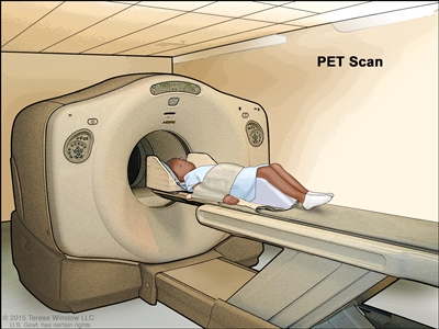
Positron emission tomography (PET) scan. The child lies on a table that slides through the PET scanner. The head rest and white strap help the child lie still. A small amount of radioactive glucose (sugar) is injected into the child's vein, and a scanner makes a picture of where the glucose is being used in the body. Cancer cells show up brighter in the picture because they take up more glucose than normal cells do. - MRI (magnetic resonance imaging): A procedure that uses a magnet, radio waves, and a computer to make a series of detailed pictures of areas inside the body. This procedure is also called nuclear magnetic resonance imaging (NMRI).
- Pulmonary function test (PFT): A test to see how well the lungs are working. It measures how much air the lungs can hold and how quickly air moves into and out of the lungs. It also measures how much oxygen is used and how much carbon dioxide is given off during breathing. This is also called a lung function test.
- Fine-needle aspiration (FNA) biopsy: The removal of tissue or fluid using a thin needle. A pathologist views the tissue or fluid under a microscope to look for cancer cells.
- Thoracoscopy: A surgical procedure to look at the organs inside the chest to check for abnormal areas. An incision (cut) is made between two ribs and a thoracoscope is inserted into the chest. A thoracoscope is a thin, tube-like instrument with a light and a lens for viewing. It may also have a tool to remove tissue or lymph node samples, which are checked under a microscope for signs of cancer. In some cases, this procedure is used to remove part of the esophagus or lung.
- Bronchoscopy: A procedure to look inside the trachea and large airways in the lung for abnormal areas. A bronchoscope is inserted through the nose or mouth into the trachea and lungs. A bronchoscope is a thin, tube-like instrument with a light and a lens for viewing. It may also have a tool to remove tissue samples, which are checked under a microscope for signs of cancer.
- Laparoscopy: A surgical procedure to look at the organs inside the abdomen to check for abnormal areas. Small incisions (cuts) are made in the wall of the abdomen and a laparoscope (thin, lighted tube) is inserted into one of the incisions. Other instruments may be inserted through the same or other incisions to perform procedures such as removing organs or taking tissue samples to be checked under a microscope for signs of cancer.
- Cytologic exam: An exam of cells under a microscope (by a pathologist) to check for anything abnormal. For mesothelioma, fluid is taken from around the lungs or from the abdomen. A pathologist checks the cells in the fluid.
Certain factors affect prognosis (chance of recovery).
Prognosis depends on whether the cancer:
- has spread throughout the thin layer of tissue or into organs.
- has just been diagnosed or has recurred (come back).