


A world-renowned pioneer and leader in minimally invasive spinal surgery, Roger Härtl, MD, continually pursues research seeking innovative and less invasive surgical and biological treatment strategies for degenerative diseases of the spine. Most recently, Dr. Härtl, Co-Director of Och Spine at NewYork-Presbyterian and Director of the Weill Cornell Medicine’s Center for Comprehensive Spine Care, is exploring novel tissue engineering techniques for the repair and regeneration of degenerated spinal discs, the most common cause of back and neck pain.
Dr. Roger Härtl
“Among the 25 nonoperative and operative physicians in our spine programs are neurosurgical spine surgeons, orthopedic spine surgeons, pain management and sports medicine specialists, and neurologists who care for some 25,000 patients with spine issues each year,” says Dr. Härtl, who is also the Hansen-MacDonald Professor of Neurological Surgery at Weill Cornell Medicine. “Many of these patients come in with disc herniations and lumbar spinal stenosis that cause structural problems, back pain, and instability. A number of these issues can be caused by a diseased disc that can then progress and cause significant pathology and pain.”
In a recent literature analysis, Dr. Härtl and his Weill Cornell Medicine colleagues reviewed 15 studies of 2,019 patients with lumbar disc herniation, evaluating their treatment outcomes. Regardless of surgical or nonoperative management, 46.2 percent of these patients experienced some degree of lower back pain long-term, lasting from 2 to 27 years, as compared to an 11.9 percent prevalence of lower back pain in the general population.
A Potential Role for Autologous Stem Cells
“Successful microdiscectomy fixes the short-term problem with discontinuation from the sciatica pain, but it doesn’t seem to affect the long-term degeneration of the disc, which can continue to progress and frequently comes back years later with other problems related to the spine. So how can you prevent that from happening?” says Dr. Härtl. “That’s why we started looking into early regenerative treatment modalities and the potential of using autologous stem cells at the time of the surgery to augment and rejuvenate the disc.”
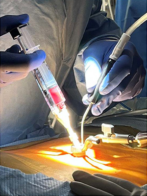
Injection of processed autologous bone marrow aspirate during lumbar microdiscectomy.
Dr. Härtl and his Weill Cornell Medicine colleagues are currently conducting a safety and feasibility prospective phase 1 clinical trial of patients undergoing microdiscectomy due to a single level, symptomatic, lumbar disc herniation. After tubular decompression of the nerve root, processed autologous bone marrow aspirate harvested from the iliac crest is injected at the disc site under fluoroscopic guidance.
“We harvest stem cells from the same patient during the surgery and then before we complete the procedure, we inject that disc with those stem cells,” says Dr. Härtl. “We are now following these patients with MRI scans and X-rays at three months and 12 months post-surgery to determine if the injection of stem cells had any effect on disc degeneration over time.”
“We’re in the process of collecting data on some 40 patients,” continues Dr. Härtl. “We haven't had any complications nor seen any negative side effects, but it’s too early to talk about huge successes. Our research is more focused on safety at this point, but obviously we’re hoping that eventually we'll see some positive results.”
Addressing the Annular Defect
“The goal of the microdiscectomy is to remove the small portion of the disc that has extruded and is now pushing on a nerve,” says Dr. Härtl. “Patients then often ask, what’s going to happen to that hole that is created by removing the disc? Are you going to repair that hole? The answer is typically no, there’s nothing that we can do right now to repair the hole. That puts every patient who has a microdiscectomy at a 10 to 20 percent risk for developing a recurrent disc herniation at the same level from the same side. This is relatively high, and patients obviously are very worried about that. And I’m concerned about that as well.”
The beauty of our approach is that it is biological and uses tissue engineering. It is very elegant and very safe if it works. And right now, it is working beautifully in animal models.
— Dr. Roger Härtl
According to Dr. Härtl, 150,000 to 200,000 patients undergo microdiscectomy each year. “If 10 percent of these patients have a recurrent disc herniation perhaps requiring more surgery and injections, it becomes a huge problem,” says Dr. Härtl, who is collaborating with Lawrence Bonassar, PhD, a biomedical engineer at Cornell University, on the development of a tissue engineered glue injected at the time of surgery to seal the annular defect.
“The glue is comprised of a collagen substance and potentially also stem cells,” says Dr. Härtl. “Our goal is annular repair and disc regeneration. We’re working on achieving FDA approval with a goal to move forward with a similar feasibility and safety study such as we’re conducting now with the autologous stem cells.”
As Dr. Härtl explains, there are currently several mechanical devices on the market that attempt to achieve the same goal. “However, there are concerns as patients don’t like the idea of having devices implanted for this purpose,” he adds. “The beauty of our approach is that it is biological and uses tissue engineering. It is very elegant and very safe if it works. And right now, it is working beautifully in animal models.”
