Jason B. Carmel, MD, PhD, Director of the Weinberg Family Cerebral Palsy Center at NewYork-Presbyterian/
Dr. Carmel directs the Movement Recovery Laboratory at NewYork-Presbyterian/
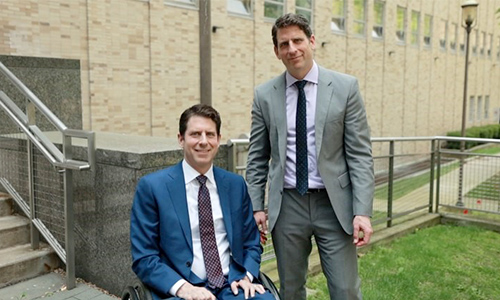
His mission is professional and also deeply personal. When Dr. Carmel was a second-year medical student at Columbia, his identical twin brother suffered a spinal cord injury, paralyzing him from the chest down and limiting the use of his hands. His brother’s injury ultimately led Dr. Carmel to become a neurologist and a neuroscientist. Now, a nerve stimulation therapy that he is developing at NewYork-Presbyterian/

Identical twins David Carmel and Dr. Jason Carmel
“Skilled movement requires motor commands and sensory feedback. We think that the pairing of brain (motor) and spinal cord (sensory) stimulation, we can strengthen their interactions in the spinal cord,” says Dr. Carmel. “The stimulation technique targets the nervous system connections spared by injury, enabling them to take over some of the lost function. Since most injuries spare some of the connections, even in people with no movement in paralyzed body parts, this is a general strategy that can be applied to many diseases and injuries.”
“Associative plasticity occurs when two stimuli converge on a common neural target,” explains Dr. Carmel, who notes early studies that targeted only the cortex to promote associative plasticity demonstrated a range of moderate effects. “However, we sought to target the strong convergence between motor and sensory systems in the spinal cord.”
Dr. Carmel and his colleagues in the Departments of Orthopedic Surgery and Neurology at NewYork-Presbyterian/
“The improvements in both function and physiology persisted for as long as they were measured, up to 50 days,” says Dr. Carmel. “The paired signals essentially mimic the normal sensory-motor integration that needs to come together to perform skilled movement. With spinal cord associative plasticity, we can precisely time the pairing of motor cortex and dorsal spinal cord stimulations to target this interaction. We think when we pair those repeatedly, we achieve better motor and sensory integration in the spinal cord. In our study, we tested the hypothesis that properly timed paired stimulation would strengthen the sensorimotor connections in the spinal cord and improve recovery after spinal cord injury. Specifically, we evaluated the physiological effects of paired stimulation, the pathways that mediate it, and its function in a preclinical trial.”
“Timing is the key,” emphasizes Dr. Carmel. “The brain and the spinal cord stimuli must arrive at the same time, which takes millisecond precision. If the stimulation is delivered to the brain and then 10 milliseconds later in the spinal cord, we get a very strong effect. If we move even 10 milliseconds on one side or the other, we lose that effect. So timing is absolutely critical for these two stimuli to arrive in the spinal cord at the same time.”
What we discovered is the importance of neural pathways to restore function. Once you know what those neural pathways are, you can then develop stimulation techniques that are specific to them just in the same way that our understanding of cholesterol biosynthesis and breakdown led to the development of specific drugs that now lower cholesterol.
— Dr. Jason Carmel
Applications in Humans
From their animal research, Dr. Carmel and the research team concluded “that spinal cord associative plasticity strengthens sensorimotor connections within the spinal cord, resulting in partial recovery of reflex modulation and forelimb function after moderate spinal cord injury. Since both motor cortex and spinal cord stimulation are performed routinely in humans, this approach can be trialled in people with spinal cord injury or other disorders that damage sensorimotor connections and impair dexterity.”
Dr. Carmel is now testing SCAP on injury patients at NewYork-Presbyterian/
“What we discovered is the importance of neural pathways to restore function,” says Dr. Carmel. “Once you know what those neural pathways are, you can then develop stimulation techniques that are specific to them just in the same way that our understanding of cholesterol biosynthesis and breakdown led to the development of specific drugs that now lower cholesterol. When it comes to spinal injuries, every little bit of function you get back could be very meaningful.”


