“We know mechanosensation is essential for oral functions, but the biology behind how these somatosensory neurons that innervate the oral cavity and upper airway contribute to orally guided functions such as flavor perception, feeding mechanics, and speech remains unexplained,” says Yalda Moayedi, PhD, Assistant Professor of Neurological Sciences in the Department of Neurology and in the Department of Otolaryngology – Head and Neck Surgery at Columbia. In fact, Dr. Moayedi’s fascination with the neural basis of somatosensation forms the basis of her research, which focuses on investigating – in humans and in animal models – the neurons and circuits that underlie oral and upper airway mechanosensation.
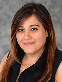
Dr. Yalda Moayedi
“Understanding the basic biology of the neurons that innervate the oral cavity and upper airway has broad implications,” explains Dr. Moayedi. “As we chew foods, we rely on central pattern generators that are modified by feedback of sensory neurons for ensuring food is kept in place. When we’re ready to swallow, mechanosensation is used to assess whether the bolus is of the proper size and salivary content to swallow without injury. When the bolus is ready, mechanosensory cues are thought to contribute to initiation and propagation of swallow.”
Dr. Moayedi notes that if the same sensory-motor systems related to feeding go awry, then dysphagia can ensue. With dysphagia affecting an estimated 15 million adults in the United States and with over 60,000 Americans dying each year from complications associated with swallowing dysfunctions, the clinical ramifications are enormous.
“The presence of a swallowing disorder causes not only quality of life issues in patients but also has significant morbidity and mortality, as dysphagia can cause aspiration pneumonia and choking,” continues Dr. Moayedi. “Despite this, we know little about the mechanosensory cells and molecules that mediate oral behaviors and airway defense. Remarkably, for such a common disorder, the primary treatment options are insufficient and often focus on changing the way you eat, physical modifications such as thickening liquids, reducing oral feeding, or changing posture. An understanding of the fundamental biology of sensory innervation is critical for developing new treatments for dysphagia and other diseases of orofacial neurons, including dysphonia and cranial neuropathies. Without this knowledge, it is difficult to create new therapeutic strategies.”
Impact of Mechanosensory Neurons Across the Lifespan
According to Dr. Moayedi, by and large the underlying mechanisms for swallowing dysfunction have been approached as motor based. “However, we know that there are sensory neurons embedded in the tissues in the mouth and throat that assess food features and signal motor neurons to initiate and modulate swallow. Additionally, recent studies have identified clinical cases of dysphagia attributed to sensory abnormalities.”
With this in mind, Dr. Moayedi noted it was reasonable to ask whether sensory dysfunction is a risk factor for dysphagia. To this end, Dr. Moayedi in collaboration with Michael J. Pitman, MD, Chief of the Division of Laryngology and the Director of the Center for Voice and Swallowing at NewYork-Presbyterian/Columbia University Irving Medical Center, Michelle S. Troche, PhD, CCC-SLP, and Ellen A. Lumpkin, PhD, carried out a study to identify the sensory neurons of the human upper airway that may be involved in swallowing and whether sensory innervation modifies with aging.
Dr. Troche is Associate Professor in the Communication Sciences and Disorders Program, Department of Biobehavioral Sciences, and Director and Principal Investigator, Laboratory for the Study of Upper Airway Dysfunction at Teachers College, Columbia University. Dr. Lumpkin, who was formerly with Columbia, is Professor of Cell Biology, Development and Physiology, Department of Molecular and Cell Biology, Helen Wills Neuroscience Institute, University of California at Berkeley.
“We undertook this project investigating innervation of the upper airway in humans and how it relates to differences in baseline swallowing function with the idea that it might prompt a susceptibility to developing swallowing disorders such as dysphagia or presbyphagia later in life,” says Dr. Moayedi.
The Columbia research team recruited volunteers, ages 20 to 80, who did not have a clinical diagnosis of dysphagia. Biopsies were taken from sites that the researchers believed would be important to swallowing, including the arytenoids, tip of the epiglottis, base of the tongue, midline posterior pharyngeal wall, and lateral pharyngeal wall. Quantitative immunohistochemistry was used to identify antibodies for all neurons, neurons that were myelinated and therefore likely to be mechanosensory, as well as chemosensory cells. Densities of lamina propria innervation, epithelial innervation, solitary chemosensory cells, and taste buds were calculated and correlated with age.
Their findings, published in the July 16, 2022 online issue of The Laryngoscope, showed:
- Arytenoid had the highest density of innervation and chemosensory cells across all measures compared to other sites
- Taste buds were frequently observed in arytenoid and epiglottis
- Base of tongue, lateral pharynx, and midline posterior pharynx had minimal innervation and few chemosensory cells
- Epithelial innervation was present primarily in close proximity to chemosensory cells and taste buds
- Overall innervation and myelinated fibers in the arytenoid lamina propria decline with aging
“We found that as humans age, they start to experience loss of specific subtypes of sensory neurons in the upper airways, particularly in the arytenoid. This is important because, in general, as people age, they are at higher risk for developing a swallowing disorder,” explains Dr. Moayedi. “In a follow-up study, we're seeing that some of the baseline differences in the sensory innervation in these upper airway sites correlate with differences in swallowing abilities, providing support for the idea that age-related alterations in sensory neurons density and morphology could impact function and be a contributing factor to presbyphagia.”
Defining Oral Sensory Neurons in the Tongue and Hard Palate
In 2021, Ardem Patapoutian, PhD, and David Julius, PhD, shared the Nobel Prize in Physiology for their groundbreaking research in the biology of senses, identifying receptors that allow the body’s cells to sense temperature and touch. Dr. Patapoutian discovered the family of mechanosensitive Piezo ion channels, including identifying the Piezo2 ion channel as the primary mediator of mechano-transcription in mammals. Dr. Julius identified a protein called TRPV1 that responds to painful heat. Their discoveries were fortuitous for Dr. Moayedi’s own quest to identify neurons in the oral cavity that mediate texture detection.
As a postdoctoral researcher in Dr. Lumpkin’s laboratory at Columbia, Dr. Moayedi analyzed the protein localization of Piezo2 in transgenic mice producing another breakthrough revelation that neurons that terminated the epithelium surrounding taste buds were found to be Piezo2-positive. Findings from this research were published in the July 2, 2018, issue of Scientific Reports. “This structure of mechanosensory ending had not been previously described in the literature and so it represented a really novel type of neuron to investigate in my lab,” says Dr. Moayedi.
Dr. Moayedi and her colleagues continued to pursue investigations of the anatomical diversity of human tongue sensory innervation. Their findings of homologous structures in the human tongues were published in the August 2021 issue of The Journal of Comparative Neurology.
“In our recently published work, we touch on the discoveries of Dr. Julius, where we show distinct patterns of TRP ion channel expression in the human tongue,” adds Dr. Moayedi. “This has helped us to better understand the oral cells and neurons that are responsible for transducing temperature, chemical compounds relevant to flavor, inflammation, and cellular stress in the mouth.”
Over the past few years, she and her colleagues have identified the anatomy of the neurons in both rodent models and humans that innervate the oral tissues. “Many senses converge in the oral cavity. We have found that in the tongue, in particular, the neurons are quite different from neurons located anywhere else in the body,” she notes. “One area that my lab is working on is trying to figure out what these different types of neurons sense, how they contribute to flavor perception, and how the tongue, with its unique mechanosensory system, is able to have the high acuity touch discrimination that it does.”
To answer these questions, Dr. Moayedi and collaborators adapted a method to record neuronal responses to mechanical stimulation of the tongue, facilitating unbiased functional classification of tongue-innervating neurons based on their physiological features. This allowed them to obtain a better understanding of the diversity and distribution of the oral mechanosensor neurons that could, in turn, guide predictions about functionality related to feeding and speech.
“Our studies are among the first to classify mechanosensory neurons innervating the tongue,” says Dr. Moayedi. “Collectively, we demonstrated for the first time to my knowledge, that tongue innervating mechanoreceptors are structurally, functionally, and molecularly distinct, and, in fact, are quite different from neurons that innervate other parts of the body. The oral surfaces are innervated by a variety of mechanosensory neurons with unique morphologies and the vast majority of tongue innervating mechanoreceptors respond best to moving stimuli. This makes sense when you think about how most tongue functions involve active movement rather than passive pressure sensing.”
Dr. Moayedi notes, “In these anatomical studies, we have identified a diverse array of putative mechanosensory endings in both the tongue and the hard palate, but what really remains unknown here is what features of stimuli do these neuron classes detect, then importantly, how do these different classes convey textural features to relay a unified version of food qualities, e.g., this chip is greasy and crispy versus compliant and dry?”
Can Mechanosensory Neurons Reveal Upper Airway Disease?
“In the context of the mouth, some neurons are tuned to sense light, sustained stimulation like the feeling of your tongue resting against your hard palate. Other neurons are tuned to detect movement facilitating detection of textures,” continues Dr. Moayedi. “The idea is that we need all of these neurons together to be able to carry out different functions, but how this relates to complicated oral behaviors is not yet well understood. Going forward, our goal is to identify contributing factors that could lead to the loss of function of mechanosensory neurons over time; preserve the function of these neurons as we age so that we can delay disorders related to loss of mechanosensation; and be able to use loss of mechanosensory function as a biomarker to detect diseases of the upper airway.”


